Color doppler
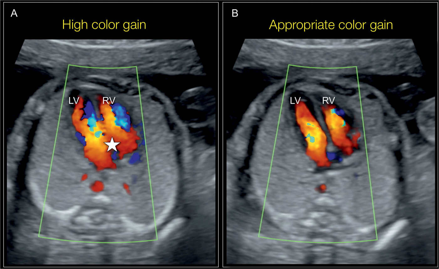
Doppler-ultrahangvizsgálat - a módszer elve, típusai és a vizsgálat .. Ezek közül egyik az úgynevezett színes (color) Doppler, ahol az érben áramló vér színkódolva látható. A vizsgálófej felé közeledő vér piros színnel látható a képernyőn, míg a távolodó kékkel. Az áramlási sebességet színárnyalatok különböztetik meg. A lassabb áramlásnál sötétebb, míg a gyorsabbnál világosabb a szín. color doppler. Color flow Doppler (ultrasound) | Radiology Reference Article .. Color flow Doppler is used frequently in sonography to semiquantitative overall blood flow to a region of interest. Depiction of the general velocity and direction of blood flow within the heart and blood vessels is of primary importance in echocardiography and vascular ultrasound respectively. color doppler. Color-doppler | KLINIKAI KÖZPONT - unideb.hu. ELLÁTÁS BETEGEKNEK, HOZZÁTARTOZÓKNAK Címlap Color-doppler. Doppler vizsgálat - Wáberer Medical Center. A Doppler, (color, duplex) vizsgálat egy rutin ultrahang diagnosztikai vizsgálattechnika. A módszer célja az erek véráramlásának, állapotának vizsgálata. A többi ultrahang-vizsgálathoz hasonlóan magas frekvenciájú ultrahangot bocsátunk a vizsgálandó testrészre, és a szövetekről visszaverődött hanghullámok mintázatait . color doppler. Color Doppler Ultrasound: Purpose, Preparation, Procedure, Results - WebMD. A Doppler ultrasound is a quick, painless way to check for problems with blood flow such as deep vein thrombosis (DVT). Find out what it is, when you need one, and how its done.. A színes doppler (color-doppler) ultrahangvizsgálatok - Radiológia. Vizsgálhatók a nyaki, a hasüregi szervek, végtagok erei is color doppler. A vizsgált személy nyakát vagy a vizsgált testrészét szabaddá teszi, levetkőzik, majd hanyatt fekszik az ágyon. Bőrét kocsonyás, az ultrahanghullámok terjedését elősegítő zselével kenik be. A vizsgálófejet ráhelyezik a bőrre, esetleg kissé rá is nyomják.. Color Doppler - Cardiovascular Education - ECG & ECHO. Color Doppler Velocities recorded in a sample volume of the pulsed wave Doppler can be presented with a color. A color scale from blue to red is conventionally used. Blue color implies velocities (movement) away from the transducer and red color implies velocities (movement) towards the transducer.. Ultrasonic colour Doppler imaging - PMC - National Center for .. Ultrasonic colour Doppler is an imaging technique that combines anatomical information derived using ultrasonic pulse-echo techniques with velocity information derived using ultrasonic Doppler techniques to generate colour-coded maps of tissue velocity superimposed on grey-scale images of tissue anatomy.. Vascular Technology Color Flow Imaging - StatPearls - NCBI Bookshelf. Color flow imaging is a vascular technology used to assess the vascular anatomy and flow within blood vessels color doppler. It relies on ultrasonographic technology to determine the flow direction, volume, and turbulence through the vessels.[1] It provides a color Doppler imaging of the relevant vasculature examined.. Doppler Ultrasound: MedlinePlus Medical Test. Color Doppler. This test uses a computer to change sound wave measurements into different colors. The colors show the speed and direction of blood flow. Power Doppler. This is a newer type of color Doppler. It can show much smaller blood vessels and slower blood flow than standard color Doppler color doppler. But it cant show the direction of blood flow.. Color Doppler | CLINICAL CENTER - unideb.hu. Doppler ultrasound can provide information on the structure of vessels (arteries and veins), as well as the blood circulation in the organs.. Doppler ultrasonography - Wikipedia. Colour Doppler shows the direction of the blood flow in red or blue (either towards or away from the transducer). Meanwhile, spectral Doppler not only shows the direction of blood flow, it also shows the phases (pulsatility) and acceleration of the blood flow.. Principles of Colour Doppler | SpringerLink. Colour Doppler was originally developed in the mid-1980s, primarily as a technique for cardiac investigation [12]. Since then its uses have expanded considerably and cover almost every aspect of the circulation. This expansion is directly related to the fact that colour Doppler represents the most comprehensive form of ultrasonic imaging .. Ultrasound Doppler: Principles, Preparation, Results and more. The main advantage of color Doppler is the fact that one can view blood flow simultaneously in many regions of the heart. However, in contrast to PW and CW Doppler, color Doppler only permits semiquantitative assessment of blood flow velocities. Color Doppler showing a mitral regurgitation (MR) and an aortic regurgitation (AR) jet.. Physical Principles of Doppler and Color Doppler Ultrasound. First Online: 04 March 2021 548 Accesses Abstract Doppler ultrasound is based on application of the Doppler effect, also called the Doppler shift. In medicine the ultrasound wave is reflected off the moving objects which are erythrocytes in blood vessels.. A nyaki erek Doppler-vizsgálata - WEBBeteg. Mikor végeznek Doppler-vizsgálatot? Nyaki Doppler- vagy color-Doppler-vizsgálatot az utóbbi időben már a komolyabb szűrések részeként is végeznek, de van néhány jellegzetes tünet vagy betegség, amelyek fennállásakor orvosa biztosan javasolni fogja egy ilyen vizsgálat elvégzését.. Basics for performing a high-quality color Doppler sonography of the .. It encodes flow direction in colors (usually blue away from the transducer and red toward it), the amplitude of mean speed by brightness, and turbulence or variance by a third color (often green). Power Doppler is a color-coded image based on Doppler-shift intensity.. Ultrahang doppler - Budai Egészségközpont - bhc.hu color doppler. Ultrahang segítségével különböző testrészek keringését tudjuk végezni beleértve az artériákat és a vénákat is color doppler
a pogány madonna teljes film indavideo
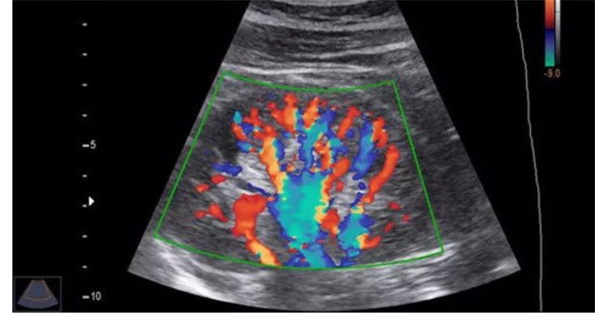
. Tájékozódni tudunk az erek faláról, tágasságáról és a keringés milyenségéről. Speciális mérések segítségével az áramlás sebességéről, ellenállás fokozódásáról vagy csökkenéséről . color doppler. PDF Doppler Ultrasound - Principles and practice - Fetal Medicine. Doppler ultrasound in general and obstetric ultrasound scanners uses pulsed wave ultrasound (Figure 4). This allows measurement of the depth (or range) of the flow site. Additionally, the size of the sample volume (or range gate) can be changed. Pulsed wave ultrasound is used to provide data for Doppler sonograms and color flow images.. Ultrahang, Vesekeringés color doppler vizsgálata - Foglaljorvost.hu. Ultrahang vizsgálat (pajzsmirigy, hasi-kismedencei, carotis doppler, ízületi, lágyrész, alsó végtag doppler vizsgálat) Uroradiológiai konzilium (két szakorvossal és kiegészítő ultrahangos vizsgálattal) Vállízületi ultrahang. Váll- ízület, izmok és inak. Végtagi artériás és vénás keringés vizsgálata.. Doppler Ultrasound: What Is It, Purpose and Procedure Details. Color Doppler: A computer changes the sound waves into different colors to show the direction of blood flow color doppler. Spectral Doppler: Graphical representation of blood flow over time. Duplex ultrasound: Combines traditional ultrasound pictures with Doppler ultrasound.. Doppler ultrasound: What is it used for? - Mayo Clinic. Doppler ultrasound is a noninvasive test that can be used to measure the blood flow through your blood vessels. It works by bouncing high-frequency sound waves off red blood cells that are circulating in the bloodstream. A regular ultrasound uses sound waves to produce images, but cant show blood flow.. What is Color Doppler Test, its Uses, Results & Normal Range?. The Colour Doppler test is a diagnostic technique that creates an image from sound waves. It provides information about the speed, movement and direction of blood flow color doppler. Your doctors will use this test to check for blockages and clots in your blood vessels. This is not possible with conventional ultrasonography as it fails to show blood flow.. Rolul ecografiei Doppler în monitorizarea pacienților cu boli venoase . color doppler. Ecografia Doppler color permite vizualizarea fluxului sangvin în culori, facilitând identificarea zonelor cu circulație slabă sau perturbată color doppler. Ecografia Doppler spectral oferă informații detaliate despre viteza și direcția fluxului sangvin, iar Doppler-ul de putere este utilizat pentru evaluarea circulației sângelui în vasele mici .. Breast ultrasound | Radiology Reference Article | Radiopaedia.org. is there a solid edge: sometimes color Doppler will help. Power Doppler and vocal fremitus. to help distinguish malignant from benign tumors. get patient to say "ahhh" or "99" very loud and observe the center of the lesion: cancer - vibrations conducted along tumor infiltration into center, hence color pixels run into center of tumor and fill it in. Ultrasonography of Superficial Soft-Tissue Masses: Society of .. Color Doppler displays the presence of flow, including mean velocity and direction. Traditional power Doppler does not show flow direction or velocity but is less dependent on the Doppler angle and is marginally more sensitive than color Doppler is to low flow color doppler. Spectral Doppler shows flow direction and patterns, including pulsatility and .. Doppler Renal Assessment, Protocols, and Interpretation. Color Doppler of the renal vein demonstrates either filling defects indicating partial occlusion or complete absence of flow consistent with obstruction color doppler. Spectral Doppler demonstrates a reversal of diastolic flow in the main renal artery, noting that, in transplant patients, a reversal of diastolic flow can also be seen with acute rejection .. Ultrasound and color Doppler applications in chronic kidney disease .. Ultrasound with color Doppler is the screening test for RAS (sensitivity and specificity of 90% and 95%, respectively) [ 24, 56, 57, 58, 58 ] color doppler. It requires a full-length sampling of renal arteries and measurement of the PSV in the ostial, medial, and hilar tracts of both arteries.. Physiologic, Histologic, and Imaging Features of Retained Products of .. Color Doppler findings include increased vascularity, predominantly in the myometrium, resulting from myometrial invasion . Figure 22 Partial mole. Photomicrograph (original magnification, ×10; H-E stain) of a suction curettage specimen obtained after spontaneous abortion at 9 weeks gestation shows a markedly hydropic villus (arrows .. Basics for performing a high-quality color Doppler sonography of the .. Color Doppler imaging. Color Doppler imaging depicts inreal-time the flow-velocity changes in the vessels color doppler
. Red and blue hues represent flow moving towards or away from the transducer. Desaturation of red or blue is an indirect measure of the velocity variations related to Doppler angle varying from 90° to 0° and 90° to 180°.. Color Doppler Flowmetry - an overview | ScienceDirect Topics. Color Doppler is a method of visually detecting motion or blood flow using a color map that is incorporated into a standard B-mode image. The principles of color Doppler are similar to those of pulsed-wave Doppler. However, a larger region can be interrogated, and detected blood flow is assigned a color, typically blue or red, depending on . color doppler. Echocardiogram: Types and What They Show - Cleveland Clinic color doppler. Doppler ultrasound. This technique shows how fast your blood flows, and also in what direction. Color Doppler ultrasound color doppler. This technique also shows your blood flow, but it uses different colors to highlight the different directions of flow color doppler. Strain imaging. This approach shows changes in how your heart muscle moves.. Ultrasound Machine Basics-Knobology, Probes, and Modes. Color Doppler Mode. The most common Doppler mode you will use is color Doppler. This mode allows you to see the movement of blood in arteries and veins with blue and red patterns on the ultrasound screen. A common question that comes up with color Doppler is: What do the colors on ultrasound mean?. Color Doppler Echocardiography - an overview - ScienceDirect. Furthermore, color Doppler echocardiography permits detection of the mitral regurgitation jet, assessment of jet direction, and evaluation of the severity of disease including jet area and vena contracta (Fig. 6). Continuous and pulse wave Doppler may be used to calculate several markers of degree of regurgitation such as EOA, regurgitant .. Ultrasound for Lower Extremity Deep Venous Thrombosis. Color Doppler ultrasound can detect complete versus incomplete obstruction. Color Doppler may help identify smaller and pelvic veins, in particular if augmentation is used. It can be used to clarify otherwise technically difficult findings color doppler. Spectral Doppler abnormalities can be used to identify obstruction in the vein segments central to the .
. Sonography Doppler Flow Imaging Instrumentation - StatPearls - NCBI .. Color Doppler is useful to interrogate organs for the presence or absence of blood flow and quickly investigate large areas for turbulent flow. In the case of cardiac imaging, it is used to qualify regurgitant and turbulent flow. Although some quantitative information can be gained by strategic use of color Doppler, it is generally useful for . color doppler. Sonography Fetal Assessment, Protocols, and Interpretation color doppler. The four-chamber view with color Doppler should show equal-sized ventricles, with defined ventricular septum, atria, and demonstration of mitral and tricuspid flow
. Compare prior imaging studies, obtain color Doppler images and possibly further imaging when applicable for accurate diagnosis.
. PDF Color Doppler Sonography Characterizing Breast Lesions color doppler. Color Doppler sonography was accessible by the 1990s, allowing a combination of grayscale and Doppler technique to effectively locate tumor vessels. Technical parameters to optimize Doppler imag-ing include a low pulsatile-repetition frequency, ideally not exceeding 1 kHz, use of appropriate. The Artifact that Tells the Truth: Color Doppler Splay Unmasking .
. The hypothetical genesis of color Doppler splay in significant MR by means of a side-lobe artifact of color Doppler ultrasound pulses. (A) While "interrogating" the imaging plane in a radial direction, pulsed Doppler side-lobe energy can encounter a jet with high Doppler power
a háború démonjai teljes film magyarul
. Color Doppler ultrasonography (CDU) provides simple, safe, noninvasive, and reproducible imaging. We therefore investigated the role of preoperative CDU combined with CTA and MRA in the quantification, typing, and diagnosis of carotid body tumors (CBTs) color doppler. We retrospectively analyzed patients with CBTs categorized into group A (type I [n = 1] and .
. Endometrial polyp | Radiology Reference Article | Radiopaedia.org. Color Doppler. feeding artery sign: a single feeding vessel may be seen extending to the polyp on color Doppler imaging 7. visualization of a vascular pedicle is 76% sensitive and 95% specific for endometrial polyps 7 color doppler. 3D ultrasound. 3D ultrasound may be useful to help delineate the borders of a polyp. color doppler. Ultrasound Evaluation of the Ovaries | Radiology Key. Color Doppler imaging ( right ) shows flow around the periphery of the mass but no flow within it. This lesion resolved on follow-up ultrasound examination. The most significant potential complication of a hemorrhagic ovarian cyst is rupture with hemoperitoneum. A ruptured hemorrhagic cyst may be difficult to distinguish from a ruptured ectopic . color doppler. Double-check Duplex Scan Documentation - AAPC Knowledge Center. Duplex ultrasound is a non-invasive evaluation of blood flow in the arteries and veins comprised of real-time images integrating B-mode, two-dimensional vascular structure, Doppler spectral analysis, and color flow Doppler imaging. Duplex scans combine conventional ultrasound with Doppler imaging color doppler. While conventional ultrasound imaging views the .. Ultrasound Assessment of Carotid Stenosis | Radiology Key. (A) Spectral Doppler image of the distal right common carotid artery ( CCA ) demonstrates a peak systolic velocity ( PSV ) of 70.5 cm/s color doppler. (B) Color Doppler image from the ipsilateral ICA shows a dense calcific plaque with posterior acoustic shadowing beyond which is an area of intense color aliasing suggesting elevated velocities.. Doppler effect - Wikipedia. The Doppler effect (also Doppler shift) is the change in the frequency of a wave in relation to an observer who is moving relative to the source of the wave. [1] [2] [3] The Doppler effect is named after the physicist Christian Doppler, who described the phenomenon in 1842 color doppler. A common example of Doppler shift is the change of pitch heard when a . color doppler. PDF American Society of Echocardiography Coding Newsletter color doppler. spectral Doppler echocardiography , and with color flow Doppler echocardiography, or 93308, Echocardiography, transthoracic, real - time with image documentation (2D), includes M-mode recording, when performed, follow-up or limited study) may also be reported. If performed, limited Doppler (93321) and color Doppler (93325) should be added services. Vscan Air™ CL - Wireless Handheld Ultrasound - GE HealthCare. Color Doppler: Color flow - color-coded overlay for real-time blood flow imaging color doppler
. PW: Pulsed wave spectral Doppler - displays speed and direction of blood flow allowing velocity measurements
. M-Mode: Motion mode - displays tissue motion over time (along one direction as indicated by M-mode cursor).